High Molecular Weight Cytokeratin P63
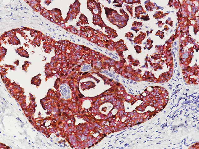
Positivity for high-molecular weight cytokeratin HMWCK. This document will discuss the potentials and pitfalls of the individual markers used in the diagnosis of prostate cancer.
Plos One Immunohistochemical Panel To Characterize Canine Prostate Carcinomas According To Aberrant P63 Expression
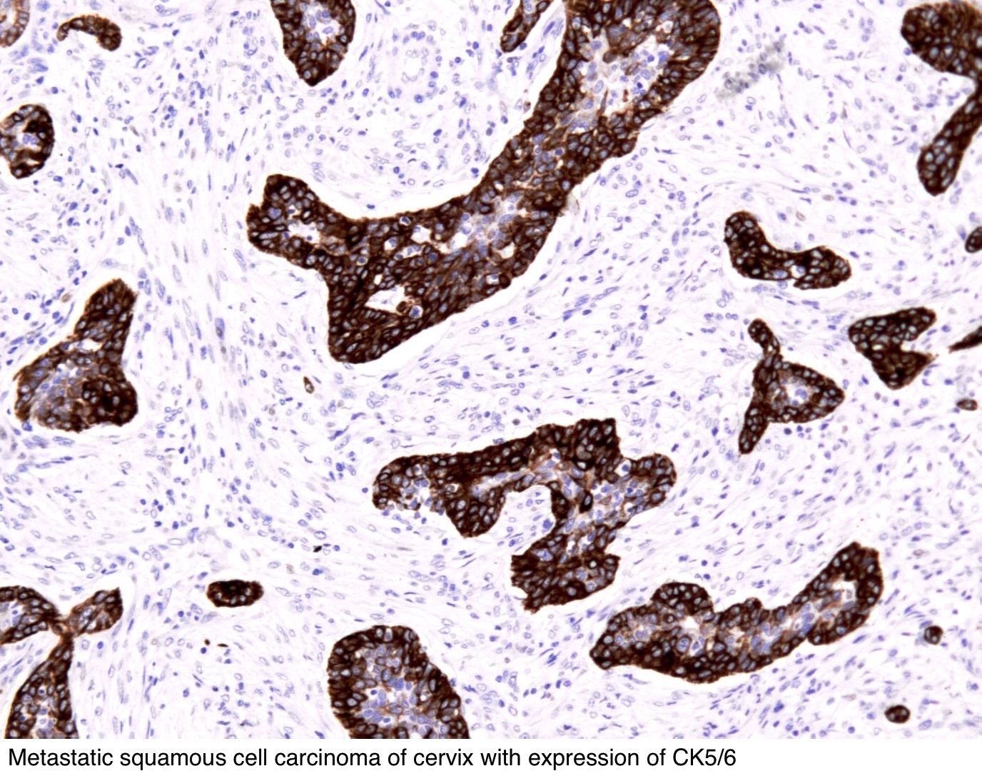
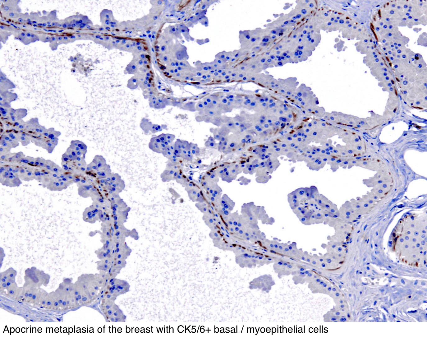
Cytokeratins CK 5 and 6 are high molecular weight basic CKs.
High molecular weight cytokeratin p63. In this study they describe another rare oc-currence of aberrant expression. Not all cases need these tests. More recently antibodies to p63 have been reported to be more sensitive than HMW-CK for the detection of prostatic basal cells.
However the antigen of HMW-CK is susceptible to the effect of formalin fixation and causes frequent loss or patchy staining in the obviously benign glands. Strong nuclear staining for p63 has been observed in benign salivary gland tumors and has also been demonstrated in BSCC and ACC. An optimal immunohistochemical panel to distinguish poorly differentiated prostate PCa from urothelial UCa carcinoma was selected from a panel consisting of prostate-specific antigen PSA and prostatic acid phosphatase PAP high-molecular-weight cytokeratin HMWCK clone 34E12 cytokeratin CK 7 CK20 p63 and -methylacyl-coenzyme A racemase.
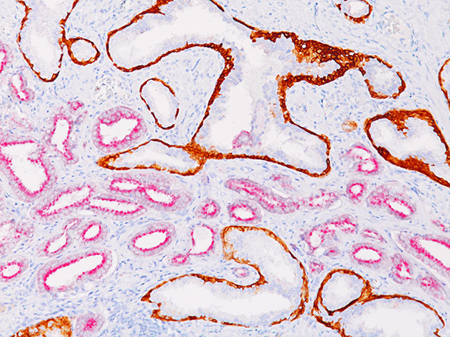
An optimal immunohistochemical panel to distinguish poorly differentiated. When carcinoma was present red cytoplasmic granular staining AMACR in the malignant glands and cells and dark brown nuclear p63 and cytoplasmic 34betaE12 staining in basal cells of adjacent. Cells may show aberrant expression for p63 immunostaining 1.
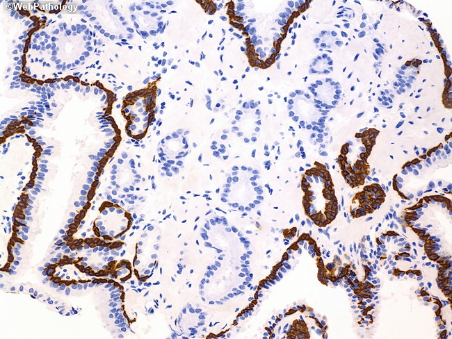
Lack of expression of high molecular weight cytoker-atin HMCK and CK7 and variable expression of RCC CD10 CA1X and PAX2. Both p63 and HMWCK are markers for basal cells which are absent in neoplastic acini. What does it mean if my biopsy report mentions special studies such as high molecular weight cytokeratin HMWCK ck903 ck56 p63 AMACR racemase 34BE12 or PIN4 cocktail.
Tumor is focally positive for PSA B 200 and negative for high- molecular-weight cytokeratin clone 34E12 C 200 and p63 D 200. All the UCs cluster together with strong diffuse reactivity for p63 predominant reactivity for CK7 and high molecular weight kinin-ogen HMWK and absent to minimal staining with PAX8 RCC antigen PAX2 alpha-methylacyl-CoA. The diagnosis of prostate adenocarcinoma is aided by IHC staining for basal cell layer markers such as p63 cytokeratin 56 CK 56 and high molecular weight cytokeratin CK HMW as well as prostate-specific markers.
PIN4 consists of a cocktail of three antibodies including AMACR P504S p63 and high molecular weight cytokeratin. The expression of p63 and highmolecularweight cytokeratins HMWCK was studied simultaneously in 33 papillary lesions including intraductal papilloma IP n 10 atypical papilloma AP n 8 and intraductal papillary carcinoma IPC n 15 by double immunostaining. We performed high molecular weight cytokeratin CK and p63 immunohistochemistry on 19 neuroendocrine carcinomas 18 basaloid squamous carcinomas and 11 solid-type adenoid cystic carcinomas.
Shiran MS 1 Tan GC Sabariah AR Rampal L Phang KS. Prostate-specific antigen high-molecular-weight cytokeratin clone 34betaE12 andor p63. Fingerprint Dive into the research topics of False positive labeling of prostate cancer with high molecular weight cytokeratin.
CK 5 is expressed by stratified squamous epithelium in a diffuse cytoplasmic pattern with perinuclear enhancement. It is a triple stain that is useful in distinguishing prostatic adenocarcinoma variable AMACRP504S red cytoplasmic staining with a lack of basal cell p63HMWK brown staining from the benign mimickers with preserved basal cell brown staining of p63HMWK and generally lack of red. E-H Poorly differentiated carcinoma with similar differential diagnosis E HE 200 resolved as poorly differentiated high-grade urothelial carcinoma.
P63 a more specific immunomarker for. In the remaining 20 of 34 cases 59 and 17 of 34 cases 50 in which the tumour cells showed strong expression of p63 and high molecular weight cytokeratin respectively larger malignant tumour cells and smaller benign basal cells of the prostatic glands and acini were highlighted with these markers and were easily distinguishable. High molecular weight keratin relatively specific for prostate basal cells Reacts to CK1 CK5 CK10 and CK14 and possibly other keratins Also called CK903 high molecular weight keratin.
These are special tests that the pathologist sometimes uses to help make the diagnosis of prostate cancer. Lesions has been High Molecular Weight-Cytokeratin HMW-CK. In recent period basal cell markers high molecular weight cytokeratin HMWCK P63 and prostate biomarker AMACR have been used as adjuvant to morphology in diagnostically challenging cases with a very high sensitivity and specificity.
P63 as a complimentary basal cell specific marker to high molecular weight-cytokeratin in distinguishing prostatic carcinoma from benign prostatic lesions. All tumors were immunostained for p63 CK 34betaE12 CK 56 synaptophysin chromogranin-A S-100 and smooth muscle actin.
 Webpathology Com A Collection Of Surgical Pathology Images
 Pathology Outlines Cytokeratin 5 6 And Ck5
 Ck Hmw P63 Amacr Rm Biocare Medical
 Pdf Prostate Specific Antigen High Molecular Weight Cytokeratin Clone 34betae12 And Or P63 An Optimal Immunohistochemical Panel To Distinguish Poorly Differentiated Prostate Adenocarcinoma From Urothelial Carcinoma
 H E Sections Top Row Of Inguinal Lymph Node With Histiocytosis Related To A Previous Hip Replacement The Histiocytes Stain Hip Replacement Lymph Nodes Stain
 Immunoperoxidase Stains At 200x Magnification A Tumor Cells Positive Download Scientific Diagram
 Pathology Outlines Cytokeratin 5 6 And Ck5
 Cmv Lymphadenitis With Prominent Monocytoid B Cell Clusters In The Subcapsular Sinus Left Arrow Cmv Inclusions Were Identified Lymph Nodes Sinusitis B Cell
 Small Focus Of Atypical Prostatic Glands In The Needle Biopsy With An Download Scientific Diagram
 Ck5 14 P63 P504s Antibody Biocare Medical
Breast Cocktail Ck Hmw P63 Ck7 8 18 Biocare Medical
 D133tp53b Is Expressed In Cancer Cells A In Situ Hybridization Using Download Scientific Diagram
 Breast Springerlink
Immunohistochemical Evaluation Of High Molecular Weight Cytokeratin Download Scientific Diagram
 Poorly Differentiated Squamous Cell Carcinoma Representative Images Of Download Scientific Diagram
 Results Of Immunohistochemical Analysis In The Present Case Download Table
 63 Expressing Prostate Tumors Are Generally Positive For Low Molecular Download Scientific Diagram
 Figure 2 Immunohistochemical Expression Of Myoepithelial Markers In Adenomyoepithelioma Of The Breast A Unique Paradoxical Staining Pattern Of High Molecular Weight Cytokeratins Springerlink
 Prostatic Adenocarcinoma With Aberrant Diffuse Expression Of High Molecular Weight Cytokeratin Pathology
Post a Comment for "High Molecular Weight Cytokeratin P63"